


How is Spinal Stenosis Diagnosed? Diagnosis, AI, and Treatment
How is spinal stenosis diagnosed? While it may feel like the answer is hidden inside your MRI, the truth is that a proper diagnosis is about much more than just a picture of your bones. In fact, imaging alone is often not that helpful.
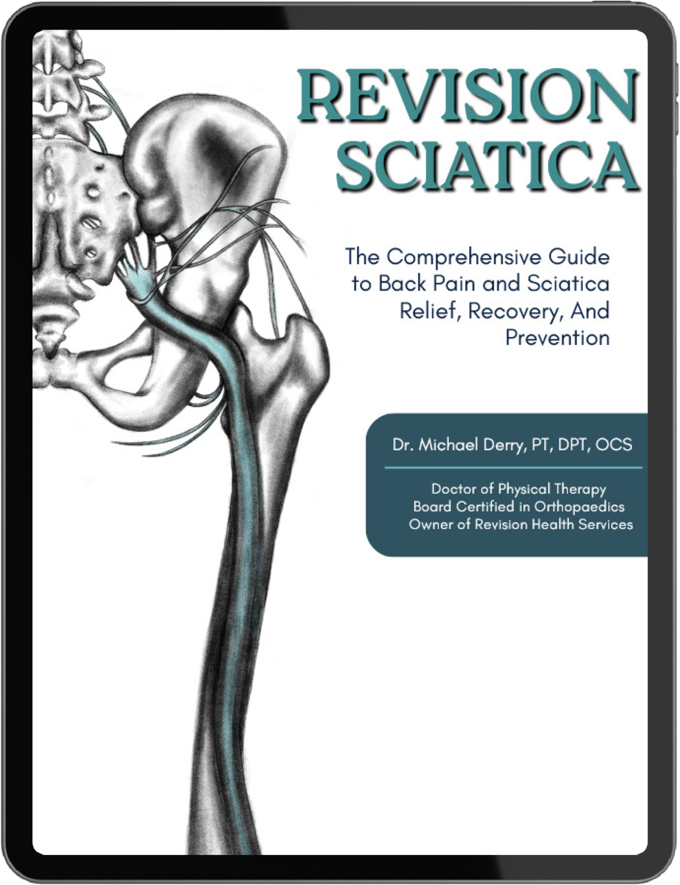
I tell every patient with stenosis: You are not your MRI. You may have significant structural changes, but they don’t have to cause pain. I often describe imaging findings as "wrinkles on the inside." Just as we expect to see wrinkles on our skin as we age, we expect to see changes in our spine. As I mentioned in my book, Revision Sciatica:
"Your spine is a robust, adaptable structure. It is not a delicate stack of blocks that is one wrong move away from collapsing. We must stop treating the image and start treating the human." — Dr. Michael Derry, DPT
The Physical Examination: Your Most Important Test
Most people think the diagnosis starts with a machine, but it actually starts with a movement screen. Stenosis is a functional diagnosis! So we rely on your symptoms and how you feel!
When I work with my clients, I am looking for the functional reality of their condition. According to the latest 2025 clinical guidelines for stenosis, a physical exam is the most reliable way to distinguish between "what they find" and "what you feel” (Meyer et al., 2025). You always treat what you feel! I can’t tell you how many times people have "awful" MRIs but feel fine.
During a physical exam, I specifically check for (and your doctor should too):
- The "Shopping Cart" Sign: As the research states, "A classic clinical sign is the 'shopping cart sign,' where patients find relief by leaning forward" (Dtsch Arztebl Int, 2025).
- Tendon Reflexes and Sensation: Since severe stenosis often involves nerve compression, we test your L4, L5, and S1 nerve roots to see if the "electrical signal" is getting through to your feet.
- Neurogenic Claudication: This is the hallmark of stenosis—heaviness or cramping that increases as you walk but is nearly gone almost immediately when you sit or bend forward.
Free Resource: If your main symptom is pain while walking, take my Spinal Stenosis Walking Pain Quiz to see if your symptoms match the classic profile of lumbar spinal stenosis.

Common Tests Your Doctor May Request
When you visit a specialist, they may request a series of tests to "map out" the narrowing. Based on current standards of care—though often not as helpful as a movement screen—here is what to expect:
- Standard MRI (Non-Contrast): The "Gold Standard" for looking at the spinal canal and nerve rootlets. It will show everything, but it may or may not match what you are feeling.
- X-rays: Often overlooked, but vital for seeing how your spine handles gravity. This helps identify spondylolisthesis (one bone sliding over another). You may have to bend forward or backward during these.
- CT Myelogram: If you have a pacemaker or cannot get an MRI, this test uses contrast dye to show exactly where the nerves are being "pinched."
- Electromyography (EMG/NCS): This test checks the actual nerves in your legs to see if there is any reduction in "flow" or electrical ability.
- Physical Therapy: Yes, physical therapy can be a test for stenosis. A clinical diagnosis is the best diagnosis, and who better to assess how you move and how your nerves and muscles communicate?
Severity Grading of Stenosis: Understanding Your Results
When your report comes back, it often uses technical grading. In the Revision Sciatica book, I explain that these grades should be used for information, not intimidation. One common system used in the literature is the Schizas Classification, which grades based on how much fluid (CSF) is left around your nerves.
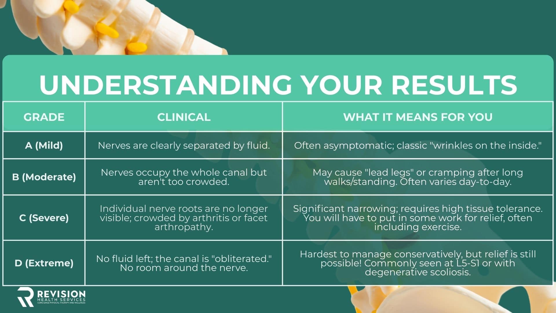
Michael’s Perspective: Research in World Neurosurgery (2025) confirms that "morphological severity on MRI does not necessarily correlate with the clinical severity of symptoms." I have seen Grade D patients walk further than Grade A patients. Your future or prognosis is determined by many more factors than the findings on your X-ray or MRI.
Learn more: You can earn better nerve health with the exercises found here: 5 Phases to Fix Lumbar Spinal Stenosis: SPARK Program.
The AI Revolution: Removing the Guesswork?
The literature today is heavily focused on the role of Artificial Intelligence (AI) in diagnosing stenosis. Human doctors often disagree on whether an MRI is "moderate" or "severe." In fact, ten radiologists reading the same MRI will often find ten different things!
As noted in Neurosurgical Focus (2024), AI is changing the game by:
- Automating Measurements: AI can calculate the "Cross-Sectional Area" (CSA) of your canal with 99% accuracy, removing human bias.
- Predictive Analytics: AI models are beginning to help us understand who will thrive with conservative care versus those who truly need surgery.
In the end, AI won’t really matter for the final diagnosis. Stenosis is a functional diagnosis. Any process that relies only on images will be limited by the machine. A computer cannot "sense" feelings or perfectly correlate a static image with your living, moving body.
From Diagnosis to Relief- What Really Works?
"In my experience, a diagnosis is just a starting point. Once we stop focusing on the 'narrowing' and start focusing on nerve health, the fear disappears and the future becomes brighter." — Dr. Michael Derry, PT, OCS
In the Revision Sciatica book, I discuss how to improve your "Nerve Tissue Tolerance." If your MRI shows narrowing, our goal is to make your nerves more resilient to that narrowing so you can feel better and do MORE without pain!
Here are the 5 phasesI typically complete with my patients to improve nerve pain, strength and more for spinal stenosis.
Take the Next Step Toward Relief
- FREE RESOURCE: Struggling with walking pain? My Spinal Stenosis Walking Pain Quiz can help you determine if your pain is likely stenosis-related.
- THE REVISION SCIATICA BOOK: Don't let a "severe" diagnosis stop you. Get the 8-week pathway that teaches you exactly how to treat your symptoms at home, including traction and TENS protocols. This includes the SPARK program, where we cover strengthening, pain relief, healing the nerve, and regaining mobility. Get the Revision Sciatica Book.
Summary
A spinal stenosis diagnosis should not rely on imaging alone—MRIs often show age-related changes that don’t necessarily cause pain. The most important part of diagnosis is a physical exam and movement assessment that evaluates how your nerves and body actually function, including signs like the “shopping cart sign,” nerve reflexes, and walking-related symptoms. Ultimately, stenosis is a functional condition, meaning treatment should focus on improving nerve health and movement rather than just reacting to what appears on a scan.
How is spinal stenosis diagnosed? While it may feel like the answer is hidden inside your MRI, the truth is that a proper diagnosis is about much more than just a picture of your bones. In fact, imaging alone is often not that helpful.
I tell every patient with stenosis: You are not your MRI. You may have significant structural changes, but they don’t have to cause pain. I often describe imaging findings as "wrinkles on the inside." Just as we expect to see wrinkles on our skin as we age, we expect to see changes in our spine. As I mentioned in my book, Revision Sciatica:
"Your spine is a robust, adaptable structure. It is not a delicate stack of blocks that is one wrong move away from collapsing. We must stop treating the image and start treating the human." — Dr. Michael Derry, DPT
The Physical Examination: Your Most Important Test
Most people think the diagnosis starts with a machine, but it actually starts with a movement screen. Stenosis is a functional diagnosis! So we rely on your symptoms and how you feel!
When I work with my clients, I am looking for the functional reality of their condition. According to the latest 2025 clinical guidelines for stenosis, a physical exam is the most reliable way to distinguish between "what they find" and "what you feel” (Meyer et al., 2025). You always treat what you feel! I can’t tell you how many times people have "awful" MRIs but feel fine.
During a physical exam, I specifically check for (and your doctor should too):
- The "Shopping Cart" Sign: As the research states, "A classic clinical sign is the 'shopping cart sign,' where patients find relief by leaning forward" (Dtsch Arztebl Int, 2025).
- Tendon Reflexes and Sensation: Since severe stenosis often involves nerve compression, we test your L4, L5, and S1 nerve roots to see if the "electrical signal" is getting through to your feet.
- Neurogenic Claudication: This is the hallmark of stenosis—heaviness or cramping that increases as you walk but is nearly gone almost immediately when you sit or bend forward.
Free Resource: If your main symptom is pain while walking, take my Spinal Stenosis Walking Pain Quiz to see if your symptoms match the classic profile of lumbar spinal stenosis.
Common Tests Your Doctor May Request
When you visit a specialist, they may request a series of tests to "map out" the narrowing. Based on current standards of care—though often not as helpful as a movement screen—here is what to expect:
- Standard MRI (Non-Contrast): The "Gold Standard" for looking at the spinal canal and nerve rootlets. It will show everything, but it may or may not match what you are feeling.
- X-rays: Often overlooked, but vital for seeing how your spine handles gravity. This helps identify spondylolisthesis (one bone sliding over another). You may have to bend forward or backward during these.
- CT Myelogram: If you have a pacemaker or cannot get an MRI, this test uses contrast dye to show exactly where the nerves are being "pinched."
- Electromyography (EMG/NCS): This test checks the actual nerves in your legs to see if there is any reduction in "flow" or electrical ability.
- Physical Therapy: Yes, physical therapy can be a test for stenosis. A clinical diagnosis is the best diagnosis, and who better to assess how you move and how your nerves and muscles communicate?
Severity Grading of Stenosis: Understanding Your Results
When your report comes back, it often uses technical grading. In the Revision Sciatica book, I explain that these grades should be used for information, not intimidation. One common system used in the literature is the Schizas Classification, which grades based on how much fluid (CSF) is left around your nerves.
Michael’s Perspective: Research in World Neurosurgery (2025) confirms that "morphological severity on MRI does not necessarily correlate with the clinical severity of symptoms." I have seen Grade D patients walk further than Grade A patients. Your future or prognosis is determined by many more factors than the findings on your X-ray or MRI.
Learn more: You can earn better nerve health with the exercises found here: 5 Phases to Fix Lumbar Spinal Stenosis: SPARK Program.
The AI Revolution: Removing the Guesswork?
The literature today is heavily focused on the role of Artificial Intelligence (AI) in diagnosing stenosis. Human doctors often disagree on whether an MRI is "moderate" or "severe." In fact, ten radiologists reading the same MRI will often find ten different things!
As noted in Neurosurgical Focus (2024), AI is changing the game by:
- Automating Measurements: AI can calculate the "Cross-Sectional Area" (CSA) of your canal with 99% accuracy, removing human bias.
- Predictive Analytics: AI models are beginning to help us understand who will thrive with conservative care versus those who truly need surgery.
In the end, AI won’t really matter for the final diagnosis. Stenosis is a functional diagnosis. Any process that relies only on images will be limited by the machine. A computer cannot "sense" feelings or perfectly correlate a static image with your living, moving body.
From Diagnosis to Relief- What Really Works?
"In my experience, a diagnosis is just a starting point. Once we stop focusing on the 'narrowing' and start focusing on nerve health, the fear disappears and the future becomes brighter." — Dr. Michael Derry, PT, OCS
In the Revision Sciatica book, I discuss how to improve your "Nerve Tissue Tolerance." If your MRI shows narrowing, our goal is to make your nerves more resilient to that narrowing so you can feel better and do MORE without pain!
Here are the 5 phasesI typically complete with my patients to improve nerve pain, strength and more for spinal stenosis.
Take the Next Step Toward Relief
- FREE RESOURCE: Struggling with walking pain? My Spinal Stenosis Walking Pain Quiz can help you determine if your pain is likely stenosis-related.
- THE REVISION SCIATICA BOOK: Don't let a "severe" diagnosis stop you. Get the 8-week pathway that teaches you exactly how to treat your symptoms at home, including traction and TENS protocols. This includes the SPARK program, where we cover strengthening, pain relief, healing the nerve, and regaining mobility. Get the Revision Sciatica Book.
Summary
A spinal stenosis diagnosis should not rely on imaging alone—MRIs often show age-related changes that don’t necessarily cause pain. The most important part of diagnosis is a physical exam and movement assessment that evaluates how your nerves and body actually function, including signs like the “shopping cart sign,” nerve reflexes, and walking-related symptoms. Ultimately, stenosis is a functional condition, meaning treatment should focus on improving nerve health and movement rather than just reacting to what appears on a scan.
%20Blog%20thumbnails.jpg)


